يعد تفسير تعداد صوره ال
 المصدر المصدر المصدردم الكامل (CBC) أحد أهم الإجراءات المختبرية
Complete Blood Count (CBC) Interpretation and Reading Results: Clinical Significance and Diagnostic Value
Abstract
Complete Blood Count (CBC) interpretation and reading results are fundamental skills in laboratory medicine because the CBC is one of the most frequently requested diagnostic tests worldwide. It provides essential information about red blood cells, white blood cells, platelets, hemoglobin, hematocrit, and red blood cell indices, allowing healthcare professionals to evaluate the patient’s overall health and detect numerous medical conditions.
The CBC is widely used to diagnose anemia, infections, inflammatory disorders, hematological malignancies, bleeding disorders, and immune system diseases. Advances in automated hematology analyzers have significantly improved the accuracy, speed, and reliability of CBC testing, making it an indispensable investigation in hospitals, emergency departments, outpatient clinics, and clinical laboratories.
Proper Complete Blood Count (CBC) interpretation and reading results require knowledge of normal reference ranges, blood cell physiology, patient history, and clinical presentation. Laboratory findings should never be interpreted in isolation but rather integrated with physical examination and additional diagnostic investigations to ensure accurate diagnosis and appropriate patient management.
This article reviews the principles of CBC testing, laboratory methodology, sample collection, major hematological parameters, clinical interpretation, common diseases, laboratory quality control, potential sources of error, and recent technological advances in hematology.
Keywords: Complete Blood Count (CBC), CBC interpretation, Hematology, Hemoglobin, Red Blood Cells, White Blood Cells, Platelets, Laboratory Diagnosis.
Introduction
Complete Blood Count (CBC) interpretation and reading results are among the most important aspects of laboratory diagnosis because the CBC provides a comprehensive assessment of the cellular components of blood. It is considered the first laboratory investigation requested for many patients presenting with symptoms such as fatigue, fever, bleeding, infection, or unexplained weakness. Due to its simplicity, affordability, and high diagnostic value, the CBC has become a cornerstone of modern clinical practice.
Blood is a specialized connective tissue responsible for transporting oxygen, nutrients, hormones, and metabolic waste products throughout the body. It also plays an essential role in immune defense, regulation of body temperature, maintenance of homeostasis, and tissue repair following injury. Since many diseases directly affect blood cells, laboratory evaluation of blood provides valuable diagnostic information for clinicians.
The Complete Blood Count measures several important hematological parameters, including the red blood cell count (RBC), white blood cell count (WBC), platelet count (PLT), hemoglobin (Hb), hematocrit (HCT), and red blood cell indices such as Mean Corpuscular Volume (MCV), Mean Corpuscular Hemoglobin (MCH), Mean Corpuscular Hemoglobin Concentration (MCHC), and Red Cell Distribution Width (RDW). Modern hematology analyzers also provide differential white blood cell counts and several advanced parameters that improve diagnostic accuracy.
Successful Complete Blood Count (CBC) interpretation and reading results require more than comparing laboratory values with normal reference ranges. Healthcare professionals must evaluate patient age, sex, medical history, medications, nutritional status, and presenting symptoms before reaching a clinical conclusion. Many abnormal laboratory findings can occur in different diseases; therefore, CBC interpretation should always be correlated with the patient’s overall clinical condition.
Recent advances in hematology technology have transformed CBC testing from manual microscopic counting into fully automated analysis using electrical impedance, flow cytometry, laser light scattering, and spectrophotometric techniques. These innovations have significantly improved laboratory efficiency while reducing analytical errors and turnaround time.
Today, the CBC remains one of the most reliable laboratory investigations available. Its ability to provide rapid, comprehensive, and clinically meaningful information makes it an indispensable diagnostic tool for disease detection, treatment monitoring, and long-term patient follow-up in healthcare institutions worldwide.
Principles of Complete Blood Count (CBC) Testing
Understanding Complete Blood Count (CBC) interpretation and reading results begins with understanding how the test is performed. The CBC is an automated laboratory investigation that evaluates the cellular components of blood using advanced hematology analyzers. Modern instruments employ several analytical technologies to ensure rapid, accurate, and reproducible measurements.
One of the primary methods used is the Coulter Principle (electrical impedance), which counts blood cells by measuring changes in electrical resistance as individual cells pass through a microscopic aperture. This technique is highly effective for determining the number and size of red blood cells and platelets.
For white blood cell analysis, many analyzers combine flow cytometry with laser light scattering. As each cell passes through a laser beam, the instrument measures scattered light from different angles to identify the size, internal structure, and complexity of the cells. This enables accurate classification of different white blood cell populations.
Hemoglobin concentration is measured using spectrophotometry. During analysis, red blood cells are lysed, releasing hemoglobin that reacts with specialized reagents. The analyzer measures light absorbance, which is directly proportional to the hemoglobin concentration.
Modern hematology analyzers also calculate additional indices such as MCV, MCH, MCHC, and RDW, providing valuable information for diagnosing different types of anemia and other hematological disorders. These technological advances have improved laboratory efficiency while reducing analytical errors and turnaround time.
Blood Sample Collection
Accurate Complete Blood Count (CBC) interpretation and reading results depend not only on analyzer performance but also on proper specimen collection and handling. Errors during the pre-analytical phase may significantly affect laboratory findings and lead to incorrect clinical decisions.
Venous blood is the preferred specimen for CBC testing. Samples are collected using standard venipuncture techniques under aseptic conditions to minimize contamination and ensure patient safety. The recommended collection container is the lavender-top tube, which contains Ethylenediaminetetraacetic Acid (EDTA) as an anticoagulant.
EDTA prevents clot formation by binding calcium ions while preserving the morphology of red blood cells, white blood cells, and platelets. Immediately after collection, the tube should be gently inverted eight to ten times to ensure complete mixing of blood with the anticoagulant. Excessive shaking should be avoided because it may cause hemolysis or platelet activation.
Samples should ideally be analyzed within a few hours after collection. Delayed analysis may alter blood cell morphology and compromise the accuracy of Complete Blood Count (CBC) interpretation and reading results. Proper specimen labeling, transportation, and storage are essential components of laboratory quality assurance.
Laboratory Equipment and Hematology Analyzer
Complete Blood Count (CBC) testing is performed using automated hematology analyzers that provide fast, accurate, and reliable measurements of blood cells. These instruments have replaced manual counting methods in most clinical laboratories because they reduce human error, improve efficiency, and increase analytical precision.
Modern hematology analyzers can process a large number of blood samples in a short period while simultaneously measuring multiple hematological parameters. In addition to counting red blood cells, white blood cells, and platelets, these analyzers calculate red blood cell indices, perform differential white blood cell counts, and generate histograms and scattergrams that assist in detecting abnormal cell populations.
Most analyzers operate using a combination of electrical impedance, flow cytometry, laser light scattering, and spectrophotometric technology. These techniques allow accurate identification of different blood cell types based on their size, internal structure, and optical properties. If abnormal findings are detected, the analyzer may generate warning flags, indicating that microscopic examination of a peripheral blood smear is required.
Several well-known manufacturers produce automated hematology analyzers, including Sysmex, Beckman Coulter, Mindray, Abbott, and Siemens Healthineers. Although each analyzer has unique features, they all follow international laboratory standards to ensure accurate and reproducible CBC results.
To maintain analytical accuracy, laboratories perform routine maintenance, instrument calibration, internal quality control, and regular performance verification. These procedures ensure that reported CBC results are reliable and suitable for clinical decision-making.
Complete Blood Count (CBC) Parameters
The Complete Blood Count includes several important laboratory parameters that provide detailed information about the cellular components of blood. Each parameter reflects a specific physiological function and contributes to the diagnosis, monitoring, and management of various medical conditions. Understanding these parameters is essential for accurate Complete Blood Count (CBC) interpretation and reading results.
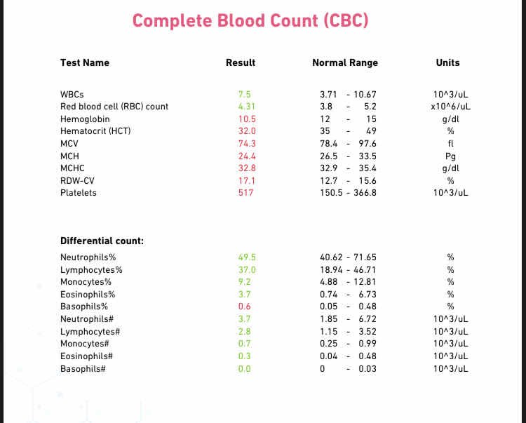
The main CBC parameters include the red blood cell count (RBC), hemoglobin (Hb), hematocrit (HCT), white blood cell count (WBC), platelet count (PLT), and red blood cell indices such as Mean Corpuscular Volume (MCV), Mean Corpuscular Hemoglobin (MCH), Mean Corpuscular Hemoglobin Concentration (MCHC), and Red Cell Distribution Width (RDW). Together, these measurements provide a comprehensive overview of the patient’s hematological status.
Red Blood Cell (RBC) Count
Red blood cells are responsible for transporting oxygen from the lungs to body tissues and returning carbon dioxide to the lungs for exhalation. This function is made possible by the presence of hemoglobin inside each red blood cell.
A decreased RBC count is commonly associated with anemia, acute blood loss, nutritional deficiencies, chronic kidney disease, or bone marrow disorders. Conversely, an increased RBC count may occur in dehydration, chronic hypoxia, smoking, high-altitude residence, or polycythemia vera.
The normal RBC count is approximately 4.7–6.1 ×10⁶/µL in adult males and 4.2–5.4 ×10⁶/µL in adult females.
Hemoglobin (Hb)
Hemoglobin (Hb) is an iron-containing protein found within red blood cells. Its primary function is to transport oxygen from the lungs to body tissues and return carbon dioxide to the lungs for exhalation. Hemoglobin is one of the most important parameters measured in a Complete Blood Count because it reflects the blood’s oxygen-carrying capacity.
Low hemoglobin levels usually indicate anemia caused by iron deficiency, vitamin B12 deficiency, folate deficiency, chronic diseases, or acute blood loss. Patients with low hemoglobin may experience fatigue, weakness, dizziness, shortness of breath, and pale skin. Conversely, elevated hemoglobin levels may occur in dehydration, chronic lung disease, smoking, or polycythemia vera.
The normal hemoglobin concentration ranges from 13.5–17.5 g/dL in adult males and 12.0–15.5 g/dL in adult females.
Hematocrit (HCT)
Hematocrit (HCT) represents the percentage of total blood volume occupied by red blood cells. It provides valuable information about the concentration of red blood cells in the bloodstream and is often interpreted together with hemoglobin and RBC count.
A low hematocrit value commonly indicates anemia, excessive blood loss, or overhydration. In contrast, a high hematocrit value may result from dehydration, chronic hypoxia, or polycythemia. Since hematocrit is influenced by both the number and size of red blood cells, it is an important parameter in evaluating various hematological conditions.
The normal hematocrit range is approximately 41–53% for adult males and 36–46% for adult females.
Mean Corpuscular Volume (MCV)
Mean Corpuscular Volume (MCV) measures the average size of red blood cells. It is one of the most useful parameters for classifying different types of anemia and guiding further laboratory investigations.
An MCV value below 80 fL indicates microcytic anemia, which is commonly caused by iron deficiency anemia or thalassemia. Values between 80 and 100 fL are considered normal and indicate normocytic red blood cells. An MCV value above 100 fL indicates macrocytic anemia, which is frequently associated with vitamin B12 or folate deficiency.
MCV is routinely interpreted together with hemoglobin, MCH, MCHC, and RDW to provide a comprehensive assessment of red blood cell abnormalities.
Mean Corpuscular Hemoglobin (MCH)
Mean Corpuscular Hemoglobin (MCH) measures the average amount of hemoglobin contained in each red blood cell. This parameter helps evaluate whether red blood cells contain adequate hemoglobin for oxygen transport.
Low MCH values are commonly seen in iron deficiency anemia and other hypochromic anemias. Elevated MCH values are usually associated with macrocytic anemia caused by vitamin B12 or folate deficiency.
The normal MCH value ranges from 27 to 33 picograms (pg) per red blood cell.
Mean Corpuscular Hemoglobin Concentration (MCHC)
Mean Corpuscular Hemoglobin Concentration (MCHC) measures the average concentration of hemoglobin within red blood cells. Unlike MCH, which measures the amount of hemoglobin, MCHC reflects how concentrated the hemoglobin is inside the cells.
Low MCHC values indicate hypochromic anemia, most commonly caused by iron deficiency. Increased MCHC values are uncommon but may occur in hereditary spherocytosis or severe red blood cell dehydration.
The normal MCHC range is 32–36 g/dL.
Red Cell Distribution Width (RDW)
Red Cell Distribution Width (RDW) measures the variation in the size of circulating red blood cells. It is useful for determining whether red blood cells are uniform in size or if there is significant variation, a condition known as anisocytosis.
An increased RDW is commonly observed in iron deficiency anemia, vitamin B12 deficiency, folate deficiency, and mixed nutritional anemias. In contrast, a normal RDW with a low MCV may suggest thalassemia trait. Therefore, RDW should always be interpreted together with MCV and other red blood cell indices to improve diagnostic accuracy.
The normal RDW value ranges between 11.5% and 14.5%.
White Blood Cell (WBC) Count
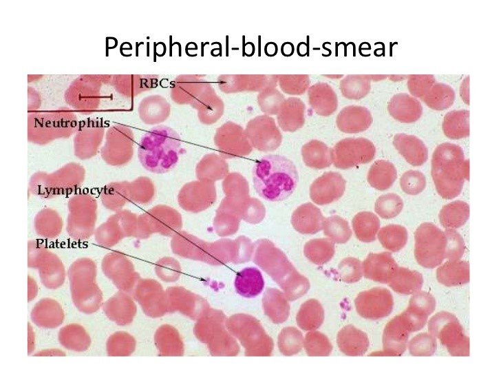
White blood cells (WBCs), also called leukocytes, are an essential part of the immune system. Their primary function is to protect the body against bacterial, viral, fungal, and parasitic infections. They also participate in inflammatory responses and immune regulation.
An elevated WBC count, known as leukocytosis, is commonly associated with bacterial infections, inflammation, physical stress, corticosteroid therapy, or hematological malignancies such as leukemia. A decreased WBC count, known as leukopenia, may occur due to viral infections, autoimmune diseases, bone marrow suppression, chemotherapy, or certain medications.
The normal white blood cell count ranges between 4,000 and 11,000 cells/µL.
White Blood Cell Differential Count
The differential white blood cell count measures the percentage of each type of white blood cell present in the circulation. This analysis provides valuable information about the body’s immune response and helps identify the underlying cause of infection or inflammation.
Neutrophils are the most abundant white blood cells and are the first line of defense against bacterial infections. Their number usually increases during acute bacterial infections.
Lymphocytes play a major role in adaptive immunity by producing antibodies and coordinating immune responses. Elevated lymphocyte counts are commonly seen in viral infections.
Monocytes remove microorganisms, damaged cells, and cellular debris through phagocytosis. Increased monocyte counts may occur in chronic infections and inflammatory disorders.
Eosinophils are primarily involved in allergic reactions and defense against parasitic infections. Elevated eosinophil counts are often associated with asthma, allergies, and parasitic diseases.
Basophils are the least common white blood cells. They release histamine and other inflammatory mediators during allergic and hypersensitivity reactions.
The differential count provides important clinical information that complements the total white blood cell count and assists physicians in establishing an accurate diagnosis.
Platelet Count (PLT)
Platelets are small, colorless cell fragments produced in the bone marrow from megakaryocytes. Their primary function is to initiate blood clot formation following blood vessel injury, thereby preventing excessive bleeding.
A decreased platelet count, known as thrombocytopenia, increases the risk of spontaneous bleeding and may result from viral infections, autoimmune diseases, bone marrow disorders, chemotherapy, or certain medications. An increased platelet count, known as thrombocytosis, may occur after inflammation, surgery, iron deficiency, or myeloproliferative disorders, increasing the risk of thrombosis.
The normal platelet count ranges between 150,000 and 450,000 platelets/µL.
Mean Platelet Volume (MPV)
Mean Platelet Volume (MPV) measures the average size of circulating platelets. Larger platelets are generally younger and more metabolically active, whereas smaller platelets are older and less active.
MPV is useful in evaluating platelet production and destruction. An increased MPV may indicate enhanced platelet production following bleeding or platelet destruction, while a decreased MPV may be associated with reduced bone marrow activity.
The normal MPV range is approximately 7.5–11.5 fL.