
Introduction
Bone
fractures are among the most common injuries affecting the musculoskeletal
system. A fracture occurs when a mechanical force exceeds the ability of the
bone to with stand it, resulting in a partial or
complete disruption of bone continuity. Fractures may occur as a result of
falls, road traffic accidents, sports injuries, direct trauma, or excessive
mechanical stress. When a fracture occurs, the damage is not limited to the
bone itself; surrounding blood vessels, bone marrow, periosteum, and soft tissues
may also be injured. This initiates a complex biological process known as fracture
healing, through which the body attempts to restore the continuity,
strength, and function of the injured bone.
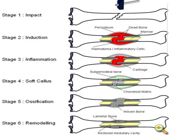
The
leg contains two long bones: the tibia and the fibula. The tibia
is the main weight-bearing bone of the lower leg, while the fibula contributes
to the stability of the ankle and provides attachment sites for muscles and
ligaments. Because of their important mechanical roles, fractures of the tibia
and fibula can significantly affect standing, walking, and normal movement.
Fracture
healing is a highly coordinated process involving inflammatory cells, blood
vessels, stem and progenitor cells, osteoblasts, osteoclasts, chondrocytes,
fibroblasts, cytokines, growth factors, and mechanical forces. Although
fracture healing is commonly divided into four major stages hematoma
formation and inflammation, soft callus formation, hard callus formation, and
bone remodeling these stages are not completely separate. Instead, they
overlap and gradually progress from one phase to another
1. Leg fracture
A
fracture is a disruption in the normal continuity of bone. The severity
of a fracture depends on the force involved, the direction of the force, the location
of the fracture, and the condition of the surrounding tissues.
Leg
fractures may involve the tibia, the fibula, or both bones. They
may be classified as closed fractures, in which the skin remains intact,
or open fractures, in which the fracture is associated with an open
wound connecting the fracture site with the external environment.
Fractures
can also differ according to their pattern, including transverse, oblique,
spiral, comminuted, and other patterns. The characteristics of the fracture
influence its stability and therefore influence the biological and mechanical
environment in which healing occurs.
Immediately
after a fracture, blood vessels within the bone and surrounding tissues are
disrupted. This results in bleeding and the accumulation of blood around the
fracture site. The resulting blood collection is known as a fracture
hematoma. The hematoma represents the initial environment in which the
subsequent inflammatory and repair processes take place.
2. Types of Fracture Healing
Fracture
healing can generally be divided into two major types: primary (direct) bone
healing and secondary (indirect) bone healing.
:Primary Bone Healing
Primary
or direct bone healing occurs when the fracture ends are accurately aligned and
there is very limited movement between them, usually because of rigid
mechanical fixation. Under these conditions, bone can heal directly across the
fracture site with relatively little external callus formation.
:Secondary Bone Healing
Secondary
or indirect bone healing is the more common form of fracture healing. It
involves the formation of a temporary callus, which gradually provides
mechanical stability and is eventually replaced and remodeled into mature bone.
Secondary
healing involves both intramembranous ossification and endochondral
ossification. The process includes inflammatory, reparative, and remodeling
events that gradually restore the structure and function of the bone.
The
following sections describe the main stages of secondary fracture healing.
3. Stage One: Hematoma Formation and
Inflammatory Phase
The
first stage of fracture healing begins immediately after the bone breaks. The
fracture damages blood vessels located within the bone, periosteum, bone
marrow, and surrounding soft tissues. As a result, bleeding occurs and blood
accumulates around the fracture site, forming a fracture hematoma.
The
hematoma contains blood cells, platelets, fibrin, and various signaling
molecules. It creates a temporary biological framework that helps organize the
subsequent healing response.
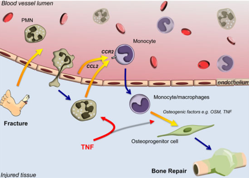
Following
the injury, the body activates an inflammatory response. Several types
of immune cells migrate to the fracture site, including neutrophils, monocytes,
and macrophages. These cells help remove damaged and dead tissue and
release signaling molecules that coordinate the healing process.
Important
inflammatory mediators include tumor necrosis factor-alpha (TNF-α), interleukin-1
(IL-1), and interleukin-6 (IL-6). These cytokines contribute to the
recruitment and activation of cells involved in tissue repair.
At
the same time, the body begins to stimulate the formation of new blood vessels
through a process known as angiogenesis. Restoration of the blood supply
is essential because newly forming tissues require oxygen, nutrients, and
cellular components supplied through the circulation.
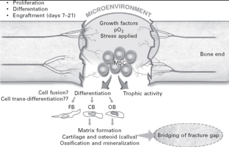
Another important event is the recruitment of mesenchymal
stem/progenitor cells (MSCs) to the fracture site. These cells can
differentiate into several cell types involved in repair, including chondrocytes,
fibroblasts, and osteoblasts
Therefore,
the inflammatory phase does not simply represent inflammation caused by injury.
It provides the biological signals and cellular environment required for the
subsequent repair phases
4. Stage Two: Soft Callus Formation
Following
the initial inflammatory response, the fracture enters the repair phase. One of
the most important events during this stage is the formation of the soft
callus, also called the fibrocartilaginous callus.
Mesenchymal
stem/progenitor cells recruited to the fracture site differentiate according to
the local biological and mechanical environment. Some cells develop into fibroblasts,
which produce connective tissue and collagen, while others differentiate into chondrocytes,
which produce cartilage.
As
these cells become active, a mixture of fibrous tissue and cartilage gradually
develops around the fracture site. This tissue forms the soft callus.
The
soft callus is not as strong as mature bone, but it provides temporary
stability and bridges the fracture gap. It also creates a biological framework
that supports the development of new bone during the next stages of healing.
The
mechanical environment is particularly important during this stage. Excessive
movement at the fracture site can interfere with normal progression of healing,
whereas an appropriate degree of mechanical stability allows the repair process
to proceed.
The
soft callus is therefore considered an intermediate structure. It represents an
important transition between the initial inflammatory stage and the formation of
stronger mineralized tissue.
During this period, vascular changes continue to
occur, and the blood supply around the fracture becomes increasingly important.
New blood vessels provide oxygen and nutrients and allow additional repair
cells to reach the area
The
soft callus gradually becomes more mineralized and is eventually replaced by
bone through processes involving endochondral ossification and direct
bone formation
5. Stage Three: Hard Callus
Formation
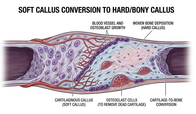
The
third major stage of fracture healing is the formation of the hard callus, also
known as the bony callus. During this stage, the relatively soft fibro
cartilaginous tissue is progressively replaced by mineralized bone.
One
of the major mechanisms involved is endochondral ossification, in which
cartilage is gradually replaced by bone. Chondrocytes undergo maturation, and
the cartilage matrix becomes mineralized. Blood vessels then grow into the
developing tissue, bringing cells and nutrients necessary for bone formation.
Osteoblasts,
which are specialized bone-forming cells, become increasingly active. They
produce an organic bone matrix called osteoid, which contains mainly type I
collagen and other proteins. The osteoid subsequently undergoes mineralization,
during which calcium and phosphate are deposited within the matrix.
An
important mineral component of bone is hydroxyapatite, a calcium-phosphate
crystal that contributes significantly to the hardness and mechanical strength
of bone.
The
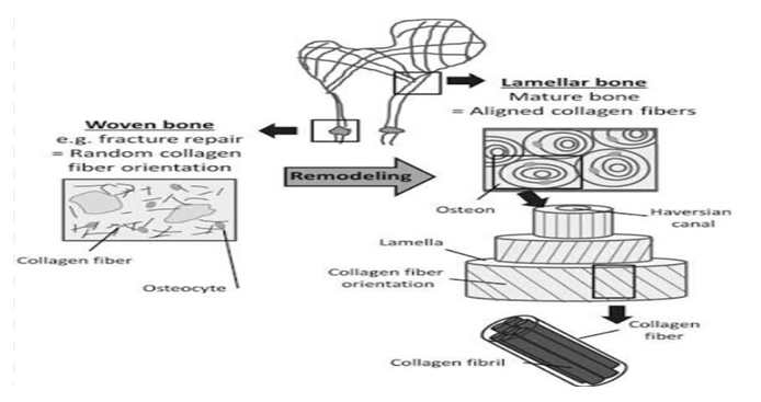
first bone produced during repair is generally called woven bone. Woven bone
has a relatively disorganized arrangement of collagen fibers compared with
mature lamellar bone. However, it provides considerably greater mechanical
stability than the soft callus.
Another
important cell type during this stage is the osteoclast. Osteoclasts are
specialized cells responsible for bone resorption, a process in which portions of
bone tissue are broken down and removed. Osteoclast activity helps shape and
reorganizes the newly formed bone.
As
hard callus formation progresses, the fracture site become increasingly stable.
New bone begins to connect the two fracture fragments, gradually restoring the
continuity of the bone.
However, the newly formed bone is not yet
identical to normal mature bone. Its structure remains relatively irregular,
which is why a further stage of healing bone remodeling is necessary.
6. Stage Four: Bone Remodeling
Bone
remodeling is the final and longest major stage of fracture healing. It begins
after sufficient new bone has formed but may continue for months or even years.
During
remodeling, the newly formed woven bone is gradually reorganized and replaced
by stronger, more mature lamellar bone. The architecture of the bone becomes
increasingly similar to its normal structure.
Two
major cell types are responsible for remodeling: osteoclasts and osteoblasts.
Osteoclasts
remove portions of the newly formed bone through bone resorption, while
osteoblasts produce new bone in a more organized pattern. The coordinated
activity of these cells allows the bone to gradually change its shape,
structure, and mechanical properties.
One
of the major objectives of remodeling is to remove excessive callus that formed
during the earlier stages. The large amount of bone initially produced around the
fracture is gradually reduced and reorganized.
The
internal structure of the bone also becomes more organized. In cortical bone,
remodeling contributes to the restoration of structures such as the Haversian
systems, which are important components of the microscopic organization of
compact bone.
Mechanical
forces also influence remodeling. Bone is a dynamic tissue that responds to the
loads placed upon it. This concept is commonly associated with Wolff’s law,
which describes the ability of bone to adapt its structure in response to
mechanical demands.
Therefore,
remodeling does not simply make the bone stronger; it also helps restore its
shape and optimize its internal architecture according to its functional
requirements.
Even after a fracture becomes clinically stable
and the patient can gradually return to normal activity, remodeling may
continue for a long period. This explains why complete biological recovery can
extend well beyond the initial period of fracture union.
7. Factors Affecting Fracture
Healing
The
rate and quality of fracture healing vary from one individual to another.
Several biological, mechanical, and environmental factors can influence the
process.
a- Blood Supply
An
adequate blood supply is essential for successful fracture healing. Blood
provides oxygen, nutrients, inflammatory cells, progenitor cells, and growth
factors required for tissue repair. Significant damage to the blood vessels
around the fracture may therefore delay healing.
b- Mechanical Stability
Mechanical
stability is another major factor. Excessive movement between fracture
fragments can interfere with the development of a stable bridge of new bone.
Appropriate stabilization helps create a mechanical environment that supports
healing.
c- Age
Age can influence bone metabolism and the speed of tissue
repair. Bone healing generally occurs more rapidly in younger individuals than
in older adults, although the exact healing process varies according to the
individual and the characteristics of the fracture.
d- Nutrition
Adequate
nutrition is necessary for tissue repair. Bone formation requires sufficient
energy, proteins, minerals, vitamins, and other nutrients. Poor nutritional
status may negatively affect the body’s ability to produce new connective and
bone tissue.
e- General Health
The
patient’s general health can also influence healing. Certain systemic diseases
and conditions may interfere with blood supply, cellular activity, metabolism,
or immune function and consequently affect fracture repair.
f- Characteristics of
the Fracture
The
location, severity, pattern, displacement, and degree of soft-tissue damage
associated with the fracture can all influence healing. Open fractures may be
particularly challenging because of the associated soft-tissue injury and
contamination risk.
g- Smoking and Other
Environmental Factors
Smoking
has been associated with impaired bone healing. Other environmental and lifestyle
factors may also influence the biological conditions required for successful
repair.
For
these reasons, fracture healing should be considered an interaction between
biological processes and the mechanical environment surrounding the fracture.
8. Healing of Tibial and Fibular
Fractures
Fractures
of the lower leg deserve particular attention because the tibia plays a
major role in weight bearing and locomotion. The characteristics of the
fracture and the condition of the surrounding soft tissues can significantly
influence healing.
The
tibia has areas where the bone is relatively close to the skin, which means that
fractures in this region may be associated with significant soft-tissue injury.
The condition of the surrounding tissues and blood supply is therefore
particularly important.
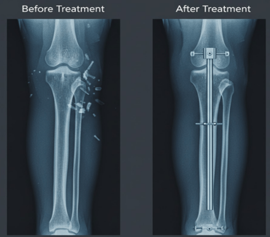
After
appropriate treatment and stabilization, the biological process of healing
continues through the stages described above. Stabilization does not itself
create new bone; rather, it provides an appropriate mechanical environment
while the body’s biological mechanisms repair the damaged tissue.
Progressive
healing can be evaluated through clinical examination and imaging techniques
such as X-ray radiography. Radiographs may demonstrate progressive
callus formation and increasing continuity across the fracture site.
However,
radiographic healing and functional recovery do not necessarily occur at
exactly the same time. A fracture may become sufficiently stable for
progressive functional activity while biological remodeling continues.
The timing of weight bearing and return to
normal activity depends on factors such as fracture type, location, treatment
method, stability, and the individual’s clinical progress. Therefore, these
decisions should be based on professional medical assessment rather than on the
appearance of the fracture alone.
9. Overall Sequence of Fracture
Healing
The
complete process of secondary fracture healing can be summarized as a sequence
of interconnected events:
Fracture
→ Hematoma formation → Inflammatory response → Recruitment of repair cells →
Soft callus formation → Hard callus formation → Woven bone formation → Bone
remodeling → Mature lamellar bone
This
sequence demonstrates that fracture healing is a dynamic process rather than a
simple formation of new bone. Inflammation prepares the injury site, the soft
callus provides temporary stability, the hard callus restores greater
mechanical strength, and remodeling reorganizes the newly formed bone into a
mature functional structure.
Importantly,
these stages overlap. For example, angiogenesis and cellular recruitment may
continue while callus formation is already occurring, and remodeling can begin
before all parts of the hard callus have completely matured.
Conclusion
Fracture
healing is a complex and highly coordinated biological process that aims to
restore the continuity, strength, structure, and function of an injured bone.
In leg fractures, particularly fractures involving the tibia and fibula,
successful healing is important for the restoration of normal weight bearing,
walking, and movement.
The
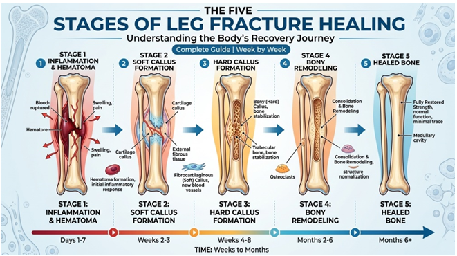
process of secondary fracture healing can be broadly divided into four major
stages. The first is hematoma formation and inflammation, during which
blood vessels are damaged, a fracture hematoma develops, and inflammatory cells
and signaling molecules are recruited to the injured area. The second stage is soft
callus formation, during which fibrous and cartilaginous tissues develop
around the fracture and provide temporary stability.
The
third stage is hard callus formation, in which the soft callus is
progressively replaced by mineralized tissue and woven bone. Osteoblasts play
an important role in producing new bone, while osteoclasts participate in the
removal and reorganization of tissue.
The
final stage is bone remodeling, during which woven bone is gradually
transformed into mature lamellar bone. Excess callus is removed, the internal
architecture of the bone becomes more organized, and the structure of the bone
adapts to its mechanical requirements.
Successful
fracture healing depends on several factors, including adequate blood supply,
appropriate mechanical stability, nutrition, age, general health, the
characteristics of the fracture, and the condition of the surrounding soft
tissues. Therefore, healing should be understood as the result of a close
interaction between biological and mechanical processes.
In conclusion, the healing of a leg fracture is
not a single event but a prolonged sequence of overlapping biological
processes. From the initial inflammatory response to the final remodeling of
mature bone, numerous cells and signaling mechanisms work together to restore
the damaged skeletal tissue and allow the bone to gradually regain its normal
function.
References
1.
Basic
concepts regarding fracture healing and the current options and future
directions in managing bone fractures.
National Center for Biotechnology Information (NCBI)/PMC.
2.
Bone
Healing and Inflammation: Principles of Fracture and Repair. National Center for Biotechnology Information (NCBI)/PMC.
3.
Inflammation,
Fracture and Bone Repair. National
Center for Biotechnology Information (NCBI)/PMC.
4.
Modulation
of the Inflammatory Response and Bone Healing. National Center for Biotechnology Information (NCBI)/PMC.
5.
Principles
of Fracture Healing and Fixation: A Literature Review. National Center for Biotechnology Information (NCBI)/PMC.
6.
American
Academy of Orthopaedic Surgeons (AAOS).
Information on fracture healing and broken bones.
1 المصدر
2 المصدر
3 المصدر
4 المصدر
