

Introduction
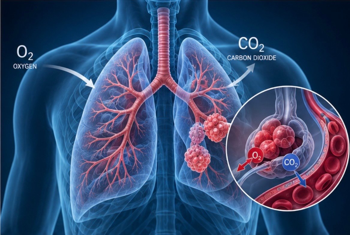
Arterial blood gas analysis, commonly called ABG, is an important laboratory test used to assess a patient’s oxygenation, ventilation, and acid–base balance. It is especially useful in critically ill patients and those with respiratory, metabolic, or circulatory problems.
An ABG measures several important values, mainly pH, PaCO₂, PaO₂, HCO₃⁻, and oxygen saturation. These values help healthcare professionals determine whether the blood is too acidic or too alkaline, whether the lungs are removing carbon dioxide properly, and whether adequate oxygen is reaching the blood.
ABG analysis is commonly used in patients with respiratory failure, severe asthma, COPD, pneumonia, sepsis, shock, diabetic ketoacidosis, kidney failure, poisoning, and other serious conditions.
1. What Is an ABG?
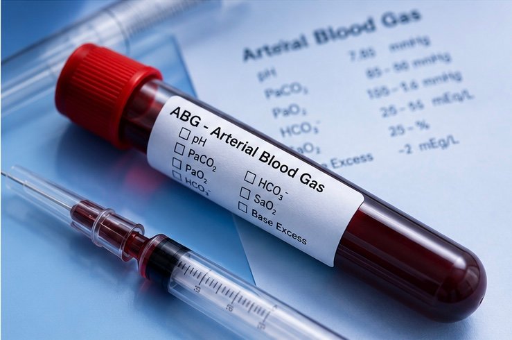
An arterial blood gas is a blood sample taken from an artery and analyzed for gases and acid–base information.
The main ABG measurements are:
- pH: Indicates how acidic or alkaline the blood is.
- PaCO₂: Indicates the amount of carbon dioxide in arterial blood and reflects ventilation.
- PaO₂: Indicates the amount of dissolved oxygen in arterial blood.
- HCO₃⁻: Represents the metabolic component of acid–base balance.
- SaO₂: Indicates the percentage of hemoglobin carrying oxygen.
- Base excess/base deficit: Gives additional information about the metabolic component.
The radial artery at the wrist is commonly used because it is relatively superficial and easy to access.
2. Why Is an ABG Performed?
ABGs are particularly useful when there is concern about:
- Respiratory failure
- Severe asthma or COPD
- Pneumonia
- Pulmonary edema
- Shock
- Sepsis
- Diabetic ketoacidosis
- Kidney failure
- Severe metabolic abnormalities
- Poisoning
- Cardiac arrest
- Patients receiving mechanical ventilation
ABGs can also be repeated to determine whether treatment is improving oxygenation, ventilation, or acid–base balance.
3. Normal ABG Values
Normal ranges can vary slightly between laboratories, but commonly used adult reference ranges are:
|
Parameter |
Approximate normal range |
|
pH |
7.35-7.45 |
|
PaCOâ‚‚ |
35-45 mmHg |
|
PaOâ‚‚ |
75-100 mmHg |
|
HCO₃⻠|
22-26 mEq/L |
|
SaOâ‚‚ |
95-100% |
|
Base excess |
Approximately-2 to +2 mEq/L |
PaO₂ must be interpreted according to factors such as age, altitude, and the amount of supplemental oxygen being given.
4. Understanding pH
The pH is the first value to examine when interpreting an ABG.
Normal arterial pH is approximately 7.35–7.45.
- pH < 7.35: Acidemia
- pH > 7.45: Alkalemia
A low pH means the blood is more acidic than normal, while a high pH means the blood is more alkaline.
The terms acidosis and alkalosis describe processes that move the body toward acidity or alkalinity. They are different from acidemia and alkalemia, which describe the actual measured blood pH.
تحليل غازات الدم الشرياني ABG وقراءته
5. PaCO₂
PaCO₂ means partial pressure of arterial carbon dioxide.
Normal PaCO₂ is approximately 35–45 mmHg.
Carbon dioxide is mainly controlled by the lungs.
- High PaCO₂ usually indicates inadequate ventilation and tends to cause respiratory acidosis.
- Low PaCO₂ usually indicates increased ventilation and tends to cause respiratory alkalosis.
For example, hypoventilation can cause carbon dioxide to accumulate, increasing PaCO₂ and lowering blood pH.
Hyperventilation causes increased removal of carbon dioxide, lowering PaCO₂ and increasing pH.
6. PaO₂
PaO₂ means partial pressure of arterial oxygen.
A typical PaO₂ on room air is approximately 75–100 mmHg, although the expected value varies with age, altitude, and other factors.
A low PaO₂ indicates hypoxemia.
Hypoxemia can occur in conditions such as:
- Pneumonia
- Pulmonary edema
- Severe asthma
- COPD
- Pulmonary embolism
- Ventilation–perfusion abnormalities
- Shunting
- Low inspired oxygen
PaO₂ should always be interpreted together with the patient’s oxygen therapy. A PaO₂ that may be acceptable on room air can be concerning if the patient is receiving a high concentration of supplemental oxygen.
تحليل غازات الدم الشرياني ABG وقراءته
7. Oxygen Saturation
SaO₂ is the percentage of hemoglobin binding sites occupied by oxygen. Normal arterial oxygen saturation is generally around 95–100%.
Pulse oximetry provides a related measurement called SpO₂.
ABG analysis can provide additional information about oxygenation that cannot always be obtained from pulse oximetry alone.
8. Bicarbonate (HCO₃⁻)
Bicarbonate is an important buffer that helps maintain normal blood pH.
Normal HCO₃⁻ is approximately 22–26 mEq/L.
- Low HCO₃⁻ commonly occurs in metabolic acidosis.
- High HCO₃⁻ commonly occurs in metabolic alkalosis.
However, bicarbonate can also change as a compensatory response to respiratory disorders. Therefore, it should always be interpreted together with pH and PaCO₂.
ABG bicarbonate is generally calculated from measured pH and PaCO₂ rather than directly measured in the same way as serum total CO₂ on a chemistry panel.
9. Base Excess and Base Deficit
Base excess provides additional information about the metabolic component of an acid–base disorder.
A negative value is commonly described as a base deficit and supports the presence of metabolic acidosis.
A positive value may support metabolic alkalosis.
The exact reference range varies between laboratories.
تحليل غازات الدم الشرياني ABG وقراءته
10. Acid–Base Balance
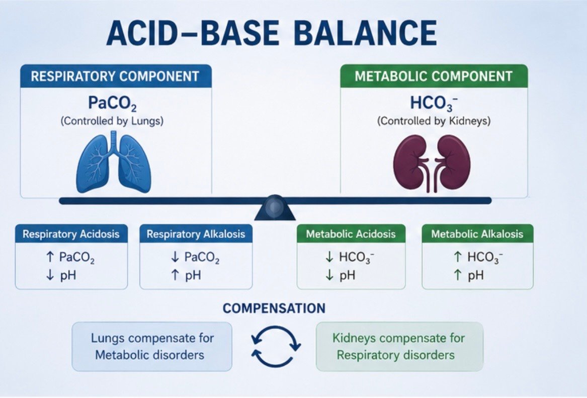
The body’s acid–base balance mainly depends on the relationship between bicarbonate and carbon dioxide.
A simple way to remember this is:
Bicarbonate = metabolic component
PaCO₂ = respiratory component
The lungs regulate carbon dioxide quickly, while the kidneys regulate bicarbonate and hydrogen ions more slowly.
Therefore:
- The lungs compensate for metabolic disorders.
- The kidneys compensate for respiratory disorders.
11. Respiratory Acidosis
Respiratory acidosis occurs when the lungs cannot remove enough carbon dioxide.
Typical ABG findings include:
- Low pH
- High PaCO₂
- HCO₃⁻ normal or increased depending on the duration
Common causes include:
- COPD exacerbation
- Severe asthma
- Airway obstruction
- Sedative or opioid effects
- Neuromuscular weakness
- Reduced respiratory drive
- Severe hypoventilation
In acute respiratory acidosis, the kidneys have not had enough time to compensate.
In chronic respiratory acidosis, the kidneys retain more bicarbonate, so HCO₃⁻ becomes higher and pH may move closer to normal.
تحليل غازات الدم الشرياني ABG وقراءته
12. Respiratory Alkalosis
Respiratory alkalosis occurs when a person removes excessive carbon dioxide through increased ventilation.
Typical findings are:
- High pH
- Low PaCO₂
- HCO₃⁻ normal or decreased depending on duration
Possible causes include:
- Anxiety or panic
- Pain
- Fever
- Pregnancy
- High altitude
- Sepsis
- Some lung diseases
- Excessive mechanical ventilation
Hyperventilation causes excessive carbon dioxide loss, resulting in a rise in pH.
13. Metabolic Acidosis
Metabolic acidosis occurs when there is excess acid, loss of bicarbonate, or both.
Typical ABG findings are:
- Low pH
- Low HCO₃⁻
- Reduced PaCO₂ due to respiratory compensation
Important causes include:
Diabetic ketoacidosis
Severe insulin deficiency causes ketone production. Ketones are acidic and can produce significant metabolic acidosis.
Lactic acidosis
Lactate can accumulate in conditions such as severe shock, sepsis, and inadequate tissue oxygen delivery.
Kidney failure
The kidneys normally remove acids and maintain bicarbonate. Severe kidney dysfunction can therefore cause metabolic acidosis.
Diarrhea
The gastrointestinal tract contains bicarbonate. Significant bicarbonate loss through diarrhea can produce metabolic acidosis.
Toxic substances
Some poisonings can produce severe metabolic acidosis.
تحليل غازات الدم الشرياني ABG وقراءته
14. Metabolic Alkalosis
Metabolic alkalosis occurs when there is excess bicarbonate or loss of acid.
Typical findings are:
- High pH
- High HCO₃⁻
- Increased PaCO₂ due to respiratory compensation
Common causes include:
- Repeated vomiting
- Gastric suction
- Certain diuretics
- Excessive acid loss
- Some hormonal disorders
Vomiting is a classic example because gastric acid is lost from the body.
15. Compensation
The body attempts to reduce changes in pH through compensation.
For a metabolic disorder, the lungs provide compensation by changing ventilation.
For a respiratory disorder, the kidneys provide compensation by changing bicarbonate and hydrogen-ion handling.
Compensation moves the pH toward normal but usually does not completely correct the original disorder.
For example, in metabolic acidosis, the patient usually breathes faster and deeper to remove CO₂.
In chronic respiratory acidosis, the kidneys retain bicarbonate to help counteract the increased CO₂.
16. Acute and Chronic Compensation
The amount of compensation depends on how long the disorder has been present.
For respiratory acidosis:
- Acute: HCO₃⁻ increases approximately 1–2 mEq/L for every 10-mmHg increase in PaCO₂.
- Chronic: HCO₃⁻ increases approximately 3–4 mEq/L for every 10-mmHg increase in PaCO₂.
For respiratory alkalosis:
- Acute: HCO₃⁻ decreases approximately 1–2 mEq/L for every 10-mmHg decrease in PaCO₂.
- Chronic: HCO₃⁻ decreases approximately 4–5 mEq/L for every 10-mmHg decrease in PaCO₂.
These are approximate clinical rules and should be interpreted with the patient’s overall condition.
تحليل غازات الدم الشرياني ABG وقراءته
17. A Simple Method for ABG Interpretation
A systematic approach makes ABG interpretation easier.
Step 1: Check the pH
- Below 7.35 → acidemia
- Above 7.45 → alkalemia
Step 2: Check PaCO₂
Ask whether carbon dioxide explains the pH.
- High PaCO₂ → respiratory acidosis
- Low PaCO₂ → respiratory alkalosis
Step 3: Check HCO₃⁻
- Low HCO₃⁻ → metabolic acidosis
- High HCO₃⁻ → metabolic alkalosis
Step 4: Identify the Primary Disorder
Determine whether the main problem is respiratory or metabolic.
Step 5: Look for Compensation
Determine whether the other system is responding appropriately.
Step 6: Assess Oxygenation
Check PaO₂ and SaO₂ and consider the patient’s oxygen therapy.
Step 7: Consider a Mixed Disorder
If the values do not fit the expected pattern, more than one acid–base disorder may be present.
تحليل غازات الدم الشرياني ABG وقراءته
18. The ROME Method
A simple learning method is ROME:
R = Respiratory Opposite
M = Metabolic Equal
In respiratory disorders, pH and PaCO₂ move in opposite directions.
Example:
pH ↓ + PaCO₂ ↑ = Respiratory acidosis
In metabolic disorders, pH and HCO₃⁻ move in the same direction.
Example:
pH ↓ + HCO₃⁻ ↓ = Metabolic acidosis
This is a useful beginner’s method, but complete interpretation also requires assessment of compensation and oxygenation.
تحليل غازات الدم الشرياني ABG وقراءته
19. Anion Gap
When metabolic acidosis is present, the anion gap can help identify the cause.
A commonly used formula is:
Anion gap = Na⁺ − (Cl⁻ + HCO₃⁻)
A high anion gap means there are additional unmeasured negatively charged substances in the blood.
Important causes include:
- Lactic acidosis
- Diabetic ketoacidosis
- Kidney failure
- Certain poisonings
Normal-anion-gap metabolic acidosis can occur when bicarbonate is lost, such as with significant diarrhea.
The anion gap should be interpreted together with electrolytes and the clinical situation.
20. Mixed Acid–Base Disorders
Sometimes a patient has more than one acid–base disorder at the same time.
For example, a patient with diabetic ketoacidosis may develop metabolic acidosis while also vomiting, which can cause metabolic alkalosis.
Mixed disorders can make the ABG appear less obvious. A normal pH does not necessarily mean that the patient’s acid–base status is normal.
Therefore, clinicians should compare the measured values with the expected compensation and consider the patient’s history and other laboratory findings.
تحليل غازات الدم الشرياني ABG وقراءته
21. ABG in Respiratory Failure
ABG analysis is especially important in respiratory failure.
Type 1 Respiratory Failure
The main problem is poor oxygenation.
PaO₂ is low, while PaCO₂ may be normal or low.
Common causes include pneumonia and pulmonary edema.
Type 2 Respiratory Failure
The main problem is inadequate ventilation and carbon dioxide retention.
PaCO₂ is elevated.
It may be associated with acute or chronic respiratory disease.
ABGs are useful for assessing both oxygenation and ventilation in these patients.
22. ABG and Mechanical Ventilation
Patients receiving mechanical ventilation may require repeated ABG testing.
ABGs can help evaluate:
- Oxygenation
- Carbon dioxide removal
- Acid–base status
- Response to treatment
For example, an elevated PaCO₂ may indicate inadequate ventilation, while a low PaO₂ may indicate inadequate oxygenation.
However, ventilator management should never be based on an ABG value alone. The patient’s clinical condition, oxygen saturation, lung function, imaging, and other information must also be considered.
23. Factors That Can Affect ABG Results
Correct sample collection and handling are important.
Potential problems include:
- Air bubbles in the sample
- Delayed analysis
- Incorrect collection technique
- Inadequate anticoagulation
- Problems with arterial-line sampling
- Incorrect patient identification
- Changes related to temperature
Samples should be handled according to the laboratory’s procedures and analyzed promptly.
24. Examples
Example 1
pH = 7.25
PaCO₂ = 55 mmHg
HCO₃⁻ = 24 mEq/L
The pH is low, indicating acidemia. PaCO₂ is high and explains the low pH. HCO₃⁻ is approximately normal.
Interpretation: Acute respiratory acidosis
Example 2
pH = 7.50
PaCO₂ = 30 mmHg
HCO₃⁻ = 23 mEq/L
The pH is high. PaCO₂ is low and explains the alkalemia.
Interpretation: Respiratory alkalosis
Example 3
pH = 7.25
PaCO₂ = 28 mmHg
HCO₃⁻ = 12 mEq/L
The pH is low and bicarbonate is low, indicating metabolic acidosis. The low PaCO₂ suggests respiratory compensation.
Interpretation: Metabolic acidosis with respiratory compensation
Example 4
pH = 7.50
PaCO₂ = 48 mmHg
HCO₃⁻ = 35 mEq/L
The pH is high and bicarbonate is high, indicating metabolic alkalosis. The elevated PaCO₂ indicates respiratory compensation.
Interpretation: Metabolic alkalosis with respiratory compensation
25. Common Mistakes
Common mistakes when interpreting ABGs include:
- Looking only at the pH.
- Ignoring PaCO₂.
- Ignoring bicarbonate.
- Forgetting compensation.
- Ignoring oxygen therapy when assessing PaO₂.
- Assuming a normal pH means the ABG is completely normal.
- Failing to consider mixed acid–base disorders.
- Interpreting the ABG without considering the patient’s clinical condition.
ABG results should always be interpreted alongside the patient’s symptoms, physical examination, oxygen requirements, electrolytes, glucose, lactate, kidney function, and other investigations.
Conclusion
Arterial blood gas analysis is an important tool for evaluating oxygenation, ventilation, and acid–base balance. The main values are pH, PaCO₂, PaO₂, HCO₃⁻, and oxygen saturation.
The easiest way to begin interpretation is to check the pH, then examine PaCO₂ and HCO₃⁻ to identify whether the main problem is respiratory or metabolic. The next step is to determine whether compensation is occurring and then assess oxygenation.
The four major acid–base disorders are respiratory acidosis, respiratory alkalosis, metabolic acidosis, and metabolic alkalosis. The lungs mainly control carbon dioxide, while the kidneys regulate bicarbonate and hydrogen ions. Compensation helps move the pH toward normal.
ABG interpretation becomes easier with practice and a consistent method. However, ABG results should never be considered in isolation. The patient’s clinical condition and other laboratory and diagnostic findings are essential for correct interpretation.
المصادر والمراجع:
- https://www.merckmanuals.com/professional/nephrology/acid-base-regulation-and-disorders/acid-base-disorders
- https://www.merckmanuals.com/professional/multimedia/table/primary-changes-and-compensations-in-simple-acid-base-disorders
- https://www.ncbi.nlm.nih.gov/books/NBK606112/
- https://www.ncbi.nlm.nih.gov/books/NBK536919/
